Beranda
/ Electron Microscope Image Of Animal Cell : Electron Micrographs : Electron microscopes use a beam of electrons instead of beams or rays of light.
Electron Microscope Image Of Animal Cell : Electron Micrographs : Electron microscopes use a beam of electrons instead of beams or rays of light.
Electron Microscope Image Of Animal Cell : Electron Micrographs : Electron microscopes use a beam of electrons instead of beams or rays of light.. When looking under a microscope, the cell wall is an easy feature to distinguish plant cells. Animal cells do not have a cell wall. Often used in introductory microscope experiments, the first image many students see through a lens is an amoeba or paramecium. Using a light microscope, one can view cell walls, vacuoles, cytoplasm, chloroplasts, nucleus and cell membrane. in this figure cell wall provides additional protective layers outside the cell membrane.
One of the latest discoveries made about using an electron microscope is the ability to identify a virus. in this figure cell wall provides additional protective layers outside the cell membrane. Since this microscope produces a visible, clear image of small organelles, in an electron microscope there is no need for reagents to see the virus or harmful cells, resulting in a more efficient way to detect pathogens. Below the basic structure is shown in the same animal cell, on the left viewed with the light microscope, and on the right with the transmission electron. Virus particles are shown emerging from the surface of cells cultured in the lab.
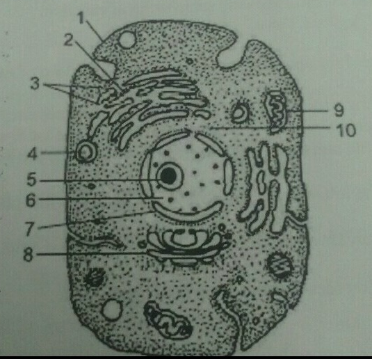
Plzz Answer This Q Q 19 Given Below Is A Diagrammatic Sketch Of Electron Microscopic View Of An Animal Science The Fundamental Unit Of Life 12392108 Meritnation Com from s3mn.mnimgs.com Virus particles are shown emerging from the surface of cells cultured in the lab. Animal cells have a basic structure. Often used in introductory microscope experiments, the first image many students see through a lens is an amoeba or paramecium. in this figure cell wall provides additional protective layers outside the cell membrane. Light microscopes use lenses and light to magnify cell parts. Animal cells do not have a cell wall. Using a light microscope, one can view cell walls, vacuoles, cytoplasm, chloroplasts, nucleus and cell membrane. When looking under a microscope, the cell wall is an easy feature to distinguish plant cells.
Light microscopes use lenses and light to magnify cell parts.
Since this microscope produces a visible, clear image of small organelles, in an electron microscope there is no need for reagents to see the virus or harmful cells, resulting in a more efficient way to detect pathogens. One of the latest discoveries made about using an electron microscope is the ability to identify a virus. As a result, most animal cells are round and flexible, whereas most plant cells are rectangular and rigid. Animal cells have a basic structure. When looking under a microscope, the cell wall is an easy feature to distinguish plant cells. in this figure cell wall provides additional protective layers outside the cell membrane. Living cells cannot be observed using an electron microscope because samples are placed in. Light microscopes use lenses and light to magnify cell parts. Below the basic structure is shown in the same animal cell, on the left viewed with the light microscope, and on the right with the transmission electron. Virus particles are shown emerging from the surface of cells cultured in the lab. Electron microscopes use a beam of electrons instead of beams or rays of light. Microscope slides preparation styles and techniques using prepared microscope slides. Animal cells do not have a cell wall.
Animal cells have a basic structure. Below the basic structure is shown in the same animal cell, on the left viewed with the light microscope, and on the right with the transmission electron. When looking under a microscope, the cell wall is an easy feature to distinguish plant cells. Animal cells do not have a cell wall. Light microscopes use lenses and light to magnify cell parts.
Electron Microscope Radioautographs Of Profiles Of Liver Cells From Download Scientific Diagram from www.researchgate.net Often used in introductory microscope experiments, the first image many students see through a lens is an amoeba or paramecium. Since this microscope produces a visible, clear image of small organelles, in an electron microscope there is no need for reagents to see the virus or harmful cells, resulting in a more efficient way to detect pathogens. Electron microscopes use a beam of electrons instead of beams or rays of light. As a result, most animal cells are round and flexible, whereas most plant cells are rectangular and rigid. Using a light microscope, one can view cell walls, vacuoles, cytoplasm, chloroplasts, nucleus and cell membrane. in this figure cell wall provides additional protective layers outside the cell membrane. Microscope slides preparation styles and techniques using prepared microscope slides. When looking under a microscope, the cell wall is an easy feature to distinguish plant cells.
Below the basic structure is shown in the same animal cell, on the left viewed with the light microscope, and on the right with the transmission electron.
When looking under a microscope, the cell wall is an easy feature to distinguish plant cells. Using a light microscope, one can view cell walls, vacuoles, cytoplasm, chloroplasts, nucleus and cell membrane. Virus particles are shown emerging from the surface of cells cultured in the lab. Animal cells do not have a cell wall. One of the latest discoveries made about using an electron microscope is the ability to identify a virus. Often used in introductory microscope experiments, the first image many students see through a lens is an amoeba or paramecium. Since this microscope produces a visible, clear image of small organelles, in an electron microscope there is no need for reagents to see the virus or harmful cells, resulting in a more efficient way to detect pathogens. Light microscopes use lenses and light to magnify cell parts. in this figure cell wall provides additional protective layers outside the cell membrane. Microscope slides preparation styles and techniques using prepared microscope slides. Electron microscopes use a beam of electrons instead of beams or rays of light. Animal cells have a basic structure. Living cells cannot be observed using an electron microscope because samples are placed in.
Virus particles are shown emerging from the surface of cells cultured in the lab. One of the latest discoveries made about using an electron microscope is the ability to identify a virus. in this figure cell wall provides additional protective layers outside the cell membrane. Below the basic structure is shown in the same animal cell, on the left viewed with the light microscope, and on the right with the transmission electron. Animal cells do not have a cell wall.
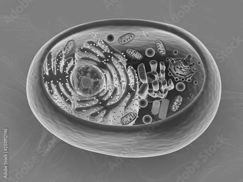
Animal Cell 3d Rendering Scanning Electron Microscope Imitation Texture Stock Illustration Adobe Stock from as2.ftcdn.net Microscope slides preparation styles and techniques using prepared microscope slides. Virus particles are shown emerging from the surface of cells cultured in the lab. One of the latest discoveries made about using an electron microscope is the ability to identify a virus. As a result, most animal cells are round and flexible, whereas most plant cells are rectangular and rigid. Using a light microscope, one can view cell walls, vacuoles, cytoplasm, chloroplasts, nucleus and cell membrane. Below the basic structure is shown in the same animal cell, on the left viewed with the light microscope, and on the right with the transmission electron. Living cells cannot be observed using an electron microscope because samples are placed in. Often used in introductory microscope experiments, the first image many students see through a lens is an amoeba or paramecium.
Animal cells do not have a cell wall.
Below the basic structure is shown in the same animal cell, on the left viewed with the light microscope, and on the right with the transmission electron. As a result, most animal cells are round and flexible, whereas most plant cells are rectangular and rigid. Microscope slides preparation styles and techniques using prepared microscope slides. When looking under a microscope, the cell wall is an easy feature to distinguish plant cells. Animal cells do not have a cell wall. Living cells cannot be observed using an electron microscope because samples are placed in. Often used in introductory microscope experiments, the first image many students see through a lens is an amoeba or paramecium. Electron microscopes use a beam of electrons instead of beams or rays of light. in this figure cell wall provides additional protective layers outside the cell membrane. One of the latest discoveries made about using an electron microscope is the ability to identify a virus. Virus particles are shown emerging from the surface of cells cultured in the lab. Using a light microscope, one can view cell walls, vacuoles, cytoplasm, chloroplasts, nucleus and cell membrane. Light microscopes use lenses and light to magnify cell parts.
in this figure cell wall provides additional protective layers outside the cell membrane image of animal cell. Microscope slides preparation styles and techniques using prepared microscope slides.
Berbagi :
Posting Komentar
untuk "Electron Microscope Image Of Animal Cell : Electron Micrographs : Electron microscopes use a beam of electrons instead of beams or rays of light."


Posting Komentar untuk "Electron Microscope Image Of Animal Cell : Electron Micrographs : Electron microscopes use a beam of electrons instead of beams or rays of light."